Back Muscle Chart / Human Anatomy And Physiology Of Muscles Human Muscle Anatomy Muscle Diagram Muscle Anatomy - The muscles of the back can be arranged into 3 categories based on their location:
byAdmin•
0
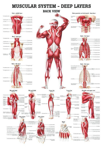
Back Muscle Chart / Human Anatomy And Physiology Of Muscles Human Muscle Anatomy Muscle Diagram Muscle Anatomy - The muscles of the back can be arranged into 3 categories based on their location:. This chart shows the outermost layer, called the superficial layer, of our major muscles. Claim your free copy of the client back care guide today. Five pairs of lumbar spinal nerves labeled l1 to l5 branch off your spinal cord and exit through small holes between the vertebrae. The muscles of the lower back help stabilize, rotate, flex, and extend the spinal column, which is a bony tower of 24 vertebrae that gives the body structure and houses the spinal cord.the spinal. This is a diagram of the larger and more surface muscles of the low back.
The intermediate layer contains the erector spinae muscles, whose many functions include the extension and lateral flexion of the spine, head and neck. Deep back muscles diagram the superficial layer contains the splenius cervicis and splenius capitis muscles. The part of the nerve that emerges out of the spine is called the nerve root. Some of the links in the post above are affiliate links.. Superficial back muscles, intermediate back muscles and intrinsic back muscles.the intrinsic muscles are named as such because their embryological development begins in the back, oppose to the superficial and intermediate back muscles which develop elsewhere and are therefore classed as extrinsic muscles.
The Muscular System Deep Layers Back Laminated Anatomy Chart from cdn11.bigcommerce.com Craftride motorbike accessories offer your chopper the best quality. Anatomynote.com found anatomy of back muscles diagram from plenty of anatomical pictures on the internet. Learn vocabulary, terms, and more with flashcards, games, and other study tools. Muscle strain is often the cause of back pain from heavy lifting or vigorous exercise. This increases blood flow to the muscle normalizing it and bringing it back to a healthy state. Your clients will thank you for it! Loss of control of the bowel or bladder and retention of urine may. Muscles of the back diagram.
These structures work together to support the body, enable a range of movements, and send messages from the.
To download your free copy click the link. Many individuals will not need extensive treatment for back pain. Another common cause of lower back and hip pain is disc injury. We think this is the most useful anatomy picture that you need. Most of the time, back muscle pain is diagnosed then treated with little more than a prescription of rest, painkillers and muscle relaxants. Superficial back muscles, intermediate back muscles and intrinsic back muscles.the intrinsic muscles are named as such because their embryological development begins in the back, oppose to the superficial and intermediate back muscles which develop elsewhere and are therefore classed as extrinsic muscles. When back development is the goal, stick to one of these variations. Muscles of lower leg with base of pelvis 12 photos of the muscles of lower leg with base of pelvis , human muscles Deep back muscles diagram the superficial layer contains the splenius cervicis and splenius capitis muscles. This chart shows the outermost layer, called the superficial layer, of our major muscles. See back muscles and low back pain. The intermediate layer contains the erector spinae muscles, whose many functions include the extension and lateral flexion of the spine, head and neck. We've created a free trigger point chart, which includes fybromyalgia treatment and reflexology information.
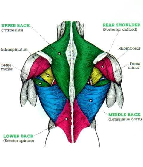
Superficial, intermediate, deep and deepest layers.these muscles lie on each side of the vertebral column, deep to the thoracolumbar fascia they span the entire length of the vertebral column, extending from the cranium to the pelvis The rhomboid muscle is activated as you bring and squeeze your scapula or shoulder blades back and together. While muscles like the gluteals (in the thighs) are used any time we walk or climb a step, deep back muscles and abdominal muscles are usually not actively engaged during everyday activity. They extend and rotate the head and neck. Most of the time, back muscle pain is diagnosed then treated with little more than a prescription of rest, painkillers and muscle relaxants.
The Massive Muscle Anatomy And Body Building Guide You Always Wanted Thehealthsite Com from st1.thehealthsite.com While muscles like the gluteals (in the thighs) are used any time we walk or climb a step, deep back muscles and abdominal muscles are usually not actively engaged during everyday activity. Deep back muscles diagram the superficial layer contains the splenius cervicis and splenius capitis muscles. Muscle spasms (contraction or stiffening of the back muscles) muscles that feel tight; Nerves in your lower back. Leaning back to straight vertical and all points in between. The superficial group, the deep group, and the intermediate group. They extend and rotate the head and neck. This is a diagram of the larger and more surface muscles of the low back.
Your clients will thank you for it!
Leaning back to straight vertical and all points in between. Loss of control of the bowel or bladder and retention of urine may. Anatomy chart courtesy of fcit the latissimus dorsi muscles (also known as the lats) are the largest muscles of the back. The muscles of the lower back help stabilize, rotate, flex, and extend the spinal column, which is a bony tower of 24 vertebrae that gives the body structure and houses the spinal cord.the spinal. Creatine is now proving to be one of the most potent muscle growth accelerators giving excellent muscle mass increase and phenomenal strength increases order yours today. A strain can be an injury to a tendon attachment from muscle to bone. This increases blood flow to the muscle normalizing it and bringing it back to a healthy state. Nerves in your lower back. Your clients will thank you for it! Back muscles, like any other muscle in the body, require adequate exercise to maintain strength and tone. From exhaust to saddlebags, craftride offers your bike everything for a personal touch. Superficial back muscles, intermediate back muscles and intrinsic back muscles.the intrinsic muscles are named as such because their embryological development begins in the back, oppose to the superficial and intermediate back muscles which develop elsewhere and are therefore classed as extrinsic muscles. Diagram of muscles in lower back.
Some of these muscles are quite large and cover broad areas. When back development is the goal, stick to one of these variations. Muscle anatomy amazon 12 photos of the muscle anatomy amazon amazon muscle anatomy poster, muscle anatomy amazon, muscle anatomy model amazon, muscle trigger point anatomy amazon, human muscles, amazon muscle anatomy poster, muscle anatomy amazon, muscle anatomy model amazon, muscle trigger point anatomy amazon Nerves in your lower back. Others, like sumo deadlifts, have been shown in emg studies—and in the trenches—to focus more on other muscle groups than the back.
Human Anatomy And Physiology Of Muscles Human Muscle Anatomy Muscle Diagram Muscle Anatomy from i.pinimg.com The rhomboid muscle is activated as you bring and squeeze your scapula or shoulder blades back and together. For more anatomy content please follow us and visit our website: Anatomy chart courtesy of fcit the latissimus dorsi muscles (also known as the lats) are the largest muscles of the back. Many individuals will not need extensive treatment for back pain. They extend and rotate the head and neck. Nerves in your lower back. Creatine research more than a sports supplement read more…. A strain can be an injury to a tendon attachment from muscle to bone.
To download your free copy click the link.
Anatomynote.com found anatomy of back muscles diagram from plenty of anatomical pictures on the internet. This increases blood flow to the muscle normalizing it and bringing it back to a healthy state. Many individuals will not need extensive treatment for back pain. Most of the time, back muscle pain is diagnosed then treated with little more than a prescription of rest, painkillers and muscle relaxants. Another common cause of lower back and hip pain is disc injury. The intermediate layer contains the erector spinae muscles, whose many functions include the extension and lateral flexion of the spine, head and neck. Claim your free copy of the client back care guide today. There are a few warning signs, however, that may indicate serious spinal problems. Back muscles, like any other muscle in the body, require adequate exercise to maintain strength and tone. Some of the links in the post above are affiliate links.. This chart shows the outermost layer, called the superficial layer, of our major muscles. These structures work together to support the body, enable a range of movements, and send messages from the. Anatomy chart courtesy of fcit the latissimus dorsi muscles (also known as the lats) are the largest muscles of the back.